

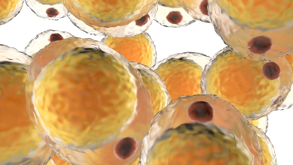
Recent research has highlighted the distinct physiological differences between subcutaneous and visceral fat, particularly concerning their response to obesity. This article will delve into the mechanisms at play, the implications of these findings, and the potential for future therapeutic interventions.
Introduction
Obesity is a growing public health concern globally, characterized by excessive fat accumulation that poses significant health risks. Notably, the location of fat deposits plays a critical role in determining the associated risks. Visceral fat, which surrounds internal organs, is more strongly correlated with metabolic dysfunction, disease prevalence, and increased mortality rates compared to subcutaneous fat found beneath the skin [1].
The Tale of Two Fats
In a groundbreaking study conducted by the German Centre for Cardiovascular Research (DZHK) and published in Nature Communications, researchers investigated the response of endothelial cells (ECs) in white adipose tissue (WAT) under conditions of diet-induced obesity [2]. The study emphasizes that fat tissue is highly vascularized, which is essential for its role in energy metabolism.
Study Design
The researchers utilized a mouse model, where subjects were fed either a normal or a high-fat diet (HFD) for eight weeks. Key findings included:
- Significant reduction in blood vessel density in both subcutaneous WAT (sWAT) and visceral WAT (vWAT), a phenomenon termed vascular rarefaction [3].
- Identification of a unique subset of endothelial cells in sWAT characterized by “fenestrae,” or pores, crucial for nutrient and hormone exchange.
- Notable downregulation of the molecular marker ITM2A in the sWAT of obese mice, indicating a loss of fenestrated ECs.
A New Cell Type and the Factor That Protects It
The researchers highlighted the role of vascular endothelial growth factor A (VEGFA) as critical for maintaining these fenestrated endothelial cells. Findings revealed that:
| Measurement | Condition | Result |
|---|---|---|
| VEGFA Levels | Obese Mice | Significant reduction in sWAT |
| VEGFA Blockade | All Mice | Reduced blood vessel density |
| VEGFA Gene Loss | Experimental Mice | Decrease in fenestrated vessels |
These observations suggest that the loss of fenestrated ECs during obesity may impair the metabolic functions of adipose tissue, contributing to obesity-related complications.
Different Responses: sWAT vs. vWAT
Interestingly, the responses of sWAT and vWAT to high-fat diet-induced obesity exhibited significant differences. The study found:
- In vWAT, ECs initiated a "vascular augmentation program," likely in an attempt to accommodate expanding tissue.
- Conversely, ECs in sWAT displayed increases in inflammation-related factors, notably the RAGE pathway.
“Our findings show that vascular changes in obesity begin earlier than previously thought and that they differ considerably depending on fat location,” said Prof. Andreas Fischer.
Implications for Future Therapies
The findings of this study illuminate new pathways in understanding and treating obesity-related complications. Potential areas of exploration include:
- Strategies to enhance blood vessel function in sWAT while managing the responses of vWAT.
- Investigating the therapeutic potential of VEGFA in regulating vascular health.
- Developing therapies tailored to the sexual dimorphism in adipose biology, as this study focused exclusively on male subjects [4].
Conclusions
In summary, as obesity rates continue to rise, understanding the complex interactions within different types of adipose tissue becomes crucial. This research underscores the importance of vascular health in the management of obesity and related disorders, paving the way for innovative therapeutic strategies.
Literature Cited
[1] Hasan, S. S., John, D., Rudnicki, M. et al. (2025). Obesity drives depot-specific vascular remodeling in male white adipose tissue. Nat Commun 16, 5392.
[2] Kuk, J. L., Katzmarzyk, P. T., Nichaman, M. Z., et al. (2006). Visceral fat is an independent predictor of all-cause mortality in men. Obesity, 14(2), 336-341.
[3] Paavonsalo, S., Hariharan, S., Lackman, M. H., & Karaman, S. (2020). Capillary rarefaction in obesity and metabolic diseases—organ-specificity and possible mechanisms. Cells, 9(12), 2683.
[4] Power, M. L., & Schulkin, J. (2008). Sex differences in fat storage, fat metabolism, and the health risks from obesity: possible evolutionary origins. British Journal of Nutrition, 99(5), 931-940.




Discussion