

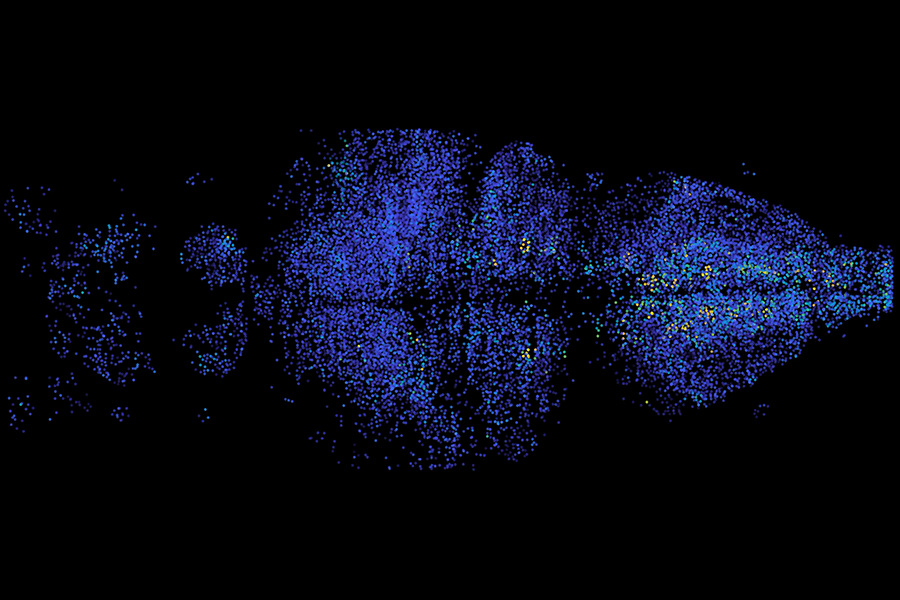
High-speed Microscopy Reveals Electrical Activity Across the Brain
Recent advances in microscopy techniques have allowed researchers to uncover the dynamic electrical activity of neurons across the brain. Utilizing high-speed volumetric imaging, scientists can now observe how neurons communicate in real time, significantly improving our understanding of brain functions involved in sensory perception, memory, and motor control.
The Technology: High-speed Microscopy
The breakthrough comes from a team led by Ed Boyden at MIT’s McGovern Institute for Brain Research. They adapted a microscopy system capable of imaging electrical activity with millisecond precision. This method enables the tracking of neural signals across the entire brain of a model organism, the zebrafish (Danio rerio). Zebrafish are particularly useful as their transparent bodies allow easy observation of brain activity.
Methodology
The key innovation in this research involves the use of genetically encoded voltage indicators (GEVIs). These are fluorescent proteins introduced into neurons, which emit light when the neuron fires. Traditional methods, such as calcium imaging, are too slow to capture the rapid electrical events of individual neurons, whereas GEVIs provide direct observation of electrical impulses.
Detailed Steps:
- Isolation of zebrafish neural tissue.
- Genetic insertion of voltage indicators into targeted neuronal populations.
- Use high-speed microscopy to capture images at 200 frames per second, allowing for the observation of rapid electrical activity.
Key Findings
The study demonstrated that:
- Neurons exhibit distinct patterns of activity in response to stimuli such as ultraviolet light.
- Activation patterns propagate throughout the brain, indicating highly coordinated neural networks. These patterns were notably observed in the optic tectum, crucial for processing visual information.
- Stimulus-independent neural activity was also detected, suggesting pre-existing networks engaged in various computations.
Significance
This new microscopy technique has immense potential to unravel complex neural networks and behaviors. By capturing how different groups of neurons coordinate in real-time, researchers can better understand the principles underlying cognitive processes and behavior.
Future Directions
The research team aims to:
- Enhance the percentage of neurons imaged.
- Improve the speed and resolution of their microscopy technique.
- Extend applications to observing brain activity in other organisms, including mice.
Limitations
While the technique offers significant advancements, challenges remain in achieving complete neuron imaging across larger brain regions and enhancing resolution without compromising speed.
Conclusion
This pioneering research lays the groundwork for a deeper understanding of how electrical impulses in neurons translate into complex behaviors and brain functions. The implications for neuroscience are profound, potentially leading to insights into neurological disorders and treatments.
References
- High-speed microscopy reveals electrical activity across the brain - MIT McGovern Institute - The technique could help scientists learn how the entire brain works to generate decisions and emotions.
- High-Speed Microscopy Unveils Brain's Electrical Activity | Mirage News - Researchers adapt a microscopy system capable of imaging electrical activity with millisecond precision.
- New microscope tracks electrical activity across the brain on the scale of milliseconds - News Medical - Explores the implications of the new high-speed imaging technique for neuroscience research.
For further reading and exploration into the implications of this study, please refer to the cited sources and related academic literature.







Discussion