


A groundbreaking study conducted by researchers at the University of Tokyo, led by Professors Takuya Yamamoto and Yasuhiro Yamada, has illuminated the intricate relationship between senescent cells and aging at the single-cell level. This research, published in the journal Nature Aging, offers invaluable insights into how these cells affect aging and the mechanisms behind their behavior in living tissues.
Understanding Cellular Senescence
Cellular senescence is characterized by a state of irreversible cell cycle arrest. It is generally triggered by factors such as:
- Oncogene activation
- DNA damage
- Oxidative stress
While senescence plays a protective role in preventing tumor development and contributes to tissue repair, its accumulation is linked to aging and the onset of various chronic diseases. The physiological roles of senescent cells have historically been challenging to elucidate due to the lack of robust in vivo models.
Innovative Mouse Models
To investigate how senescent cells behave within living tissues, the researchers engineered two mouse models with inducible expressions of:
- Constitutively active MEK1 (caMEK1)
- Constitutively active MKK6 (caMKK6)
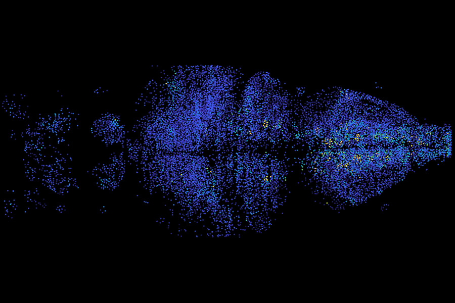
These models activate the ERK and p38 MAPK signaling pathways, both of which are known to trigger senescence in vitro. Coupled with a dual-color labeling system that differentiates between primary senescent cells (marked by red fluorescence) and secondary senescent cells (marked by green fluorescence), the researchers were able to analyze gene expression at the single-cell resolution and trace senescence propagation across tissues.
Key Findings from Transcriptomic Analyses
The study unearthed several crucial findings regarding the characteristics and behavior of senescent cells. Below is a summary of the significant observations:
| Observation | Description |
|---|---|
| Hallmark Features of Senescence | Increased p21 expression, DNA damage responses, and SASP observed in liver and colon tissues. |
| Heterogeneity of Profiles | Senescent cells exhibited variable transcriptomic profiles depending on tissue type and location. |
| Secondary Senescence Induction | SASP factors like IL-1β were found to induce secondary senescence in surrounding cells. |
The Impact of Senescence on Tissue Function
One of the most striking findings of this research was how senescence disrupted liver zonation, a crucial spatial organization for liver function. This disruption resulted in:
| Effect | Description |
|---|---|
| Impaired Metabolism | Reduced expression of metabolic genes similar to changes seen in aged tissues. |
| Comparative Transcriptomics | Transcriptomic profiles of senescent cells in mouse models mirrored those found in aged humans. |
Significance and Future Directions
This comprehensive in vivo characterization of senescent cells sheds light on cellular aging mechanisms and sets the stage for future investigations. Notably, the research emphasizes the role of intercellular signaling, particularly through SASP and Notch pathways, in driving secondary senescence and contributing to tissue dysfunction.
“Our findings highlight the diverse functions of senescent cells within tissues and establish foundational knowledge for developing therapies to target these cells and mitigate their harmful effects on aging.” – Professor Takuya Yamamoto
The caMEK1 and caMKK6 mouse models provide powerful tools for advancing our understanding of aging at the organismal level, with potential implications for therapeutic strategies aimed at managing age-related diseases.
References
Yuko Sogabe et al. (2025). Characterizing primary and secondary senescence in vivo. Nature Aging. DOI: 10.1038/s43587-025-00917-y.
For further reading, visit the article here.






Discussion