

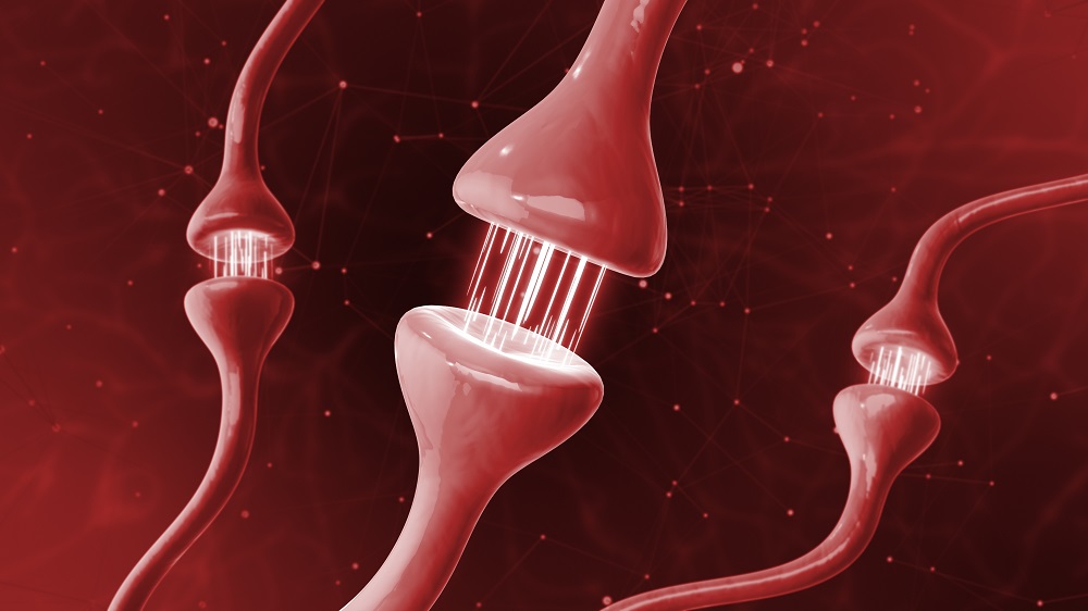
A recent study has provided critical insights into the detrimental impact of senescent astrocytes on neuronal health and synaptic formation, suggesting that the decline in certain signaling molecules may exacerbate cognitive decline in the aging brain.
Introduction
The role of astrocytes, a type of glial cell in the brain, has drawn considerable attention due to their essential functions in supporting and modulating neuronal activity. Healthy astrocytes are known to promote synaptic growth through the secretion of various factors, including thrombospondin-1 (TSP-1). However, a decline in TSP-1 production in senescent astrocytes correlates with decreased synaptic formation and potential cognitive deficits.
Significance of Senescence
Cellular senescence is widely recognized as a contributing factor to neurodegeneration and cognitive impairment. The SAMP8 mouse model, which exhibits accelerated senescence, serves as a valuable tool for understanding these processes. This research highlights the adverse effects of astrocytic senescence on neural health, particularly its influence on synapses:
- Increased senescence markers: The study showed that astrocytes from SAMP8 mice exhibited heightened levels of SA-β-gal, a marker of senescence.
- Impact on synaptic growth: Conditional media derived from senescent astrocytes hindered the ability of neural stem cells to form new synapses, reinforcing the link between astrocytic health and neuronal function.
Research Findings
The investigation focused on the conditioned media (CM) from senescent astrocytes, revealing notable differences when compared to control astrocytes. The following key observations were made:
| Aspect of Study | Control Group (Non-Senescent) | SAMP8 Group (Senescent) |
|---|---|---|
| SA-β-gal Expression | Low | High |
| Synapse Formation in CM | High | Significantly Reduced |
| TSP-1 Protein Levels | Normal | Decreased |
These results emphasize the critical role of TSP-1 in maintaining synaptic health. As previous studies have indicated, TSP-1 levels decline with age, reflecting a broader decline in factors crucial for cognitive maintenance.
Investigating TSP-1
Researchers conducted further experiments to explore the role of TSP-1 in this process:
- Use of Gabapentin: Administration of gabapentin, which inhibits TSP-1 signaling, nullified the differences in synaptic growth between CM derived from SAMP8 and control astrocytes.
- Restoring TSP-1 Levels: The introduction of TSP-1 into the conditioned media from SAMP8 astrocytes significantly enhanced synapse formation.
- Overexpression Studies: Engineering SAMP8 astrocytes to overexpress the _Thbs1_ gene, which encodes TSP-1, also resulted in improved synaptic capabilities.
Future Directions
Given these findings, researchers aim to further investigate whether TSP-1 can be utilized as a therapeutic agent to ameliorate cognitive decline associated with aging. Potential approaches include:
- Conducting in vivo studies to determine the efficacy of TSP-1 treatment in naturally aged rodents.
- Examining cognitive function alongside neuroplasticity restoration in models engineered to overexpress _Thbs1_.
Conclusion
As the landscape of neurodegenerative disease research expands, the role of senescent astrocytes and their signaling molecules, such as TSP-1, emerges as a promising avenue for investigation. The potential to restore neuroplasticity and cognitive function through targeted interventions highlights the importance of continued research in this area.
Literature Cited
[1] Akiguchi, I., et al. (2017). SAMP8 mice as a neuropathological model of accelerated brain aging and dementia: Toshio Takeda’s legacy and future directions. Neuropathology, 37(4), 293-305.
[2] Phatnani, H., & Maniatis, T. (2015). Astrocytes in neurodegenerative disease. Cold Spring Harbor Perspectives in Biology, 7(6), a020628.
[3] Clarke, L. E., et al. (2018). Normal aging induces A1-like astrocyte reactivity. Proceedings of the National Academy of Sciences, 115(8), E1896-E1905.
[4] Cheng, C., et al. (2016). Astrocyte-secreted thrombospondin-1 modulates synapse and spine defects in the fragile X mouse model. Molecular Brain, 9(1), 74.
About the Author
Josh Conway
Josh has been writing and editing articles for Lifespan over the past decade and is responsible for the continued production of daily news content. He has a programming background and is a long-time supporter of anti-aging medicine.
We would like to ask you a small favor. We are a non-profit foundation, and unlike some other organizations, we have no shareholders and no products to sell you. All our news and educational content is free for everyone to read, but it does mean that we rely on the help of people like you. Every contribution, no matter if it’s big or small, supports independent journalism and sustains our future.
© 2025 Lifespan Research Institute – ALL RIGHTS RESERVED









Discussion