

Mitochondria Delivery Method Rescues Parkinson’s in Mice
This approach reduces symptoms of Leigh and Parkinson's in these animals.
Date: March 27, 2026
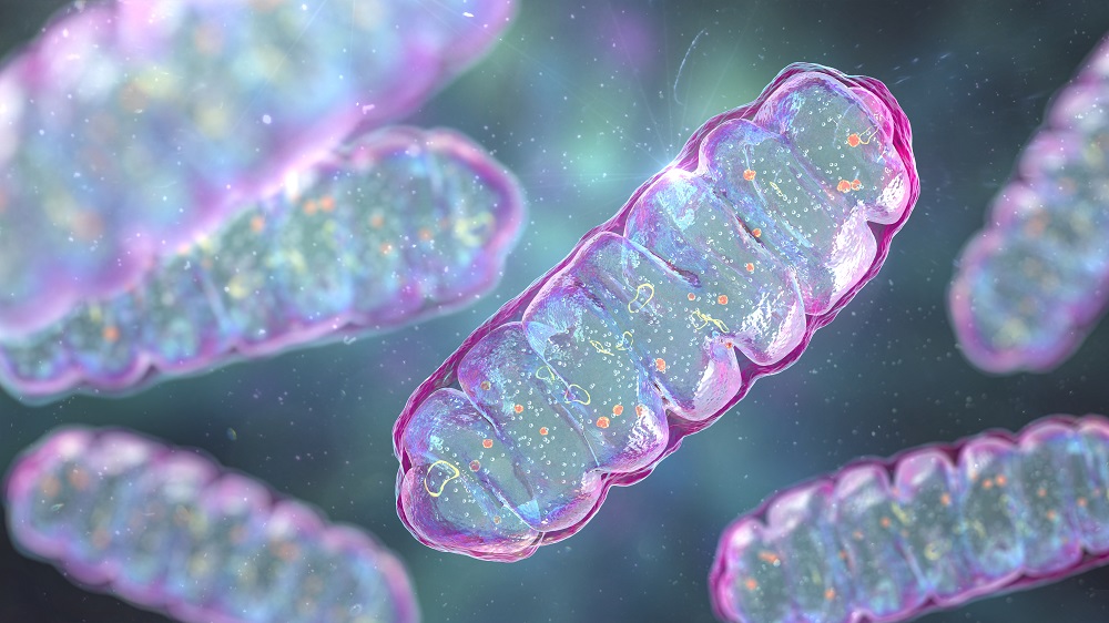
Mitochondria, encapsulated and placed into the bloodstream or target tissues, can be taken up by recipient cells. This alleviates mitochondrial diseases in cells and in animals. Scientists used red blood cells as membrane donors to encapsulate healthy mitochondria and send them into diseased cells [1].
The Delivery Problem
Mitochondrial diseases represent a heterogeneous group of disorders arising from mitochondrial malfunctions, commonly caused by mutations in mitochondrial DNA (mtDNA) or in nuclear genes responsible for encoding proteins associated with mitochondrial function. Such dysfunctions can significantly contribute to the aging process.
Given that mitochondria serve as the primary source of energy for most cellular functions, their impairments—stemming from mutations or damage due to oxidative stress—pose serious threats to cellular health. Parkinson’s disease exemplifies a neurodegenerative condition in which mitochondrial dysfunction plays a pivotal role [2].
Despite the potential benefits of delivering healthy, functional mitochondria to diseased cells, researchers have faced various obstacles. While physical methods like optical tweezers provide precision in transferring mitochondria, they are limited to only a small number of cells. On the other hand, simply injecting free mitochondria into the bloodstream has yielded only modest effects [3].
Success in a Dish
In a recent study published in Cell, a team of Chinese scientists sought to overcome these challenges by encapsulating healthy mitochondria within cellular membranes derived from red blood cells (erythrocytes). This innovative approach aims to protect mitochondria during circulation and promote their uptake by targeted cells.
Using erythrocyte plasma membranes devoid of organelles ensures their clinical safety as a membrane source. The researchers isolated mitochondria from donor cells, mixed them with erythrocyte membranes, and allowed self-assembly into capsule-like structures. Notably, mitochondria enclosed within the capsules exhibited enhanced markers of function, including increased membrane potential and ATP levels, compared to free mitochondria, indicating that encapsulation may preserve mitochondrial integrity.
Time-lapse videos demonstrated successful fusion of the capsules with recipient cells. Remarkably, within 48 hours, ~80% of acceptor cells acquired donor mitochondria, merging them into their existing mitochondrial networks. Transplanted mitochondria maintained normal membrane potential levels, with donor mtDNA constituting 71% of the total mtDNA pool.
Experiments with Cells Lacking mtDNA
The research team proceeded to test rho zero (ρ0) cells—cells lacking all mtDNA—demonstrating that donor mitochondria could significantly improve mitochondrial function and morphology. The study findings illustrated a recovery from dysfunctional swollen forms to normal tubular shapes and saw mtDNA levels restored to near-normal states persisting for at least 21 days, with evidence of active reading and translation of mtDNA-encoded genes.
Next, GM04516 human fibroblasts with deletions in mtDNA were treated using mitochondria capsules from normal human fibroblasts. Impressively, 86% of the patient cells incorporated donor mitochondria, lowering the fraction of deleted mtDNA from 14.4% to 2.67%. Enhanced oxygen consumption, ATP production, and overall cell viability were also noted.
In further tests, fibroblasts harboring the m.3243A>G mutation—a notable pathogenic mtDNA mutation—showed decreased mutation rates from 92.6% to 73.3% after treatment, along with increased mitochondrial protein levels.
Improvements in Leigh and Parkinson’s Models
With promising in vitro results, the researchers then proceeded to in vivo experiments, administering the mitochondrial capsules through various routes, including intramuscular, direct brain injection, and intravenous (IV) pathways. They also extended their trials to cynomolgus monkeys.
Transplanted mitochondria were detected in muscle tissue following intramuscular injection, the substantia nigra and cortex post-direct brain injection, and were distributed throughout the body following IV injection. Notably, in cynomolgus monkeys, mitochondrial delivery to muscle tissue was successfully achieved.
Effects in Mice with Leigh Syndrome
The application of the mitochondrial capsules significantly affected mice exhibiting Leigh syndrome, a rare and often fatal mitochondrial disorder. Median survival increased markedly from 48.5 days (untreated) to 74 days with the encapsulated mitochondrial treatment.
Effects in Parkinson's Disease Models
In the mouse model of Parkinson’s disease, which was induced by a toxin causing mitochondrial dysfunction in dopaminergic neurons, IV injections of mitochondrial capsules administered twice a week for one month led to a notable rescue of functioning dopaminergic neurons and a marked reversal of bradykinesia. The treatment enhanced mitochondrial function, with effects lasting up to three months post-therapy.
Intriguingly, a single intracerebral injection into the substantia nigra produced abundant delivery of transplanted mitochondria and marked recovery in neuronal function, further underscoring the effectiveness of targeted mitochondrial delivery.
Conclusion
As we ponder the implications of these findings, it is essential to acknowledge that many hurdles remain. However, the potential for mitochondria delivery methods to revolutionize interventions in mitochondrial diseases and neurodegenerative disorders presents a thrilling prospect for future research and clinical applications.
We would like to ask you a small favor. We are a non-profit foundation, and unlike some other organizations, we have no shareholders and no products to sell you. All our news and educational content is free for everyone to read, but it does mean that we rely on the help of people like you. Every contribution, no matter if it’s big or small, supports independent journalism and sustains our future.
Literature
- Du, S., Long, Q., Zhou, Y., Fu, J., Wu, H., Yang, L., … & Liu, X. (2026). . Cell.
- Schapira, A. H. V., Cooper, J. M., Dexter, D., Clark, J. B., Jenner, P., & Marsden, C. D. (1990). . Journal of neurochemistry, 54(3), 823-827.
- Nakai, R., Varnum, S., Field, R. L., Shi, H., Giwa, R., Jia, W., … & Brestoff, J. R. (2024). . Nature metabolism, 6(10), 1886-1896.
About the Author
Arkadi Mazin
Arkadi is a seasoned journalist and op-ed author with a passion for learning and exploration. His interests span from politics to science and philosophy. Having studied economics and international relations, he is particularly interested in the social aspects of longevity and life extension. He strongly believes that life extension is an achievable and noble goal that has yet to take its rightful place on the very top of our civilization’s agenda – a situation he is eager to change.
Support Our Mission
104% Funded
News and Topics
Receive Our Newsletter
Sign up to stay informed about the latest news and opportunities with LRI!
© 2025 Lifespan Research Institute – ALL RIGHTS RESERVED




Discussion