

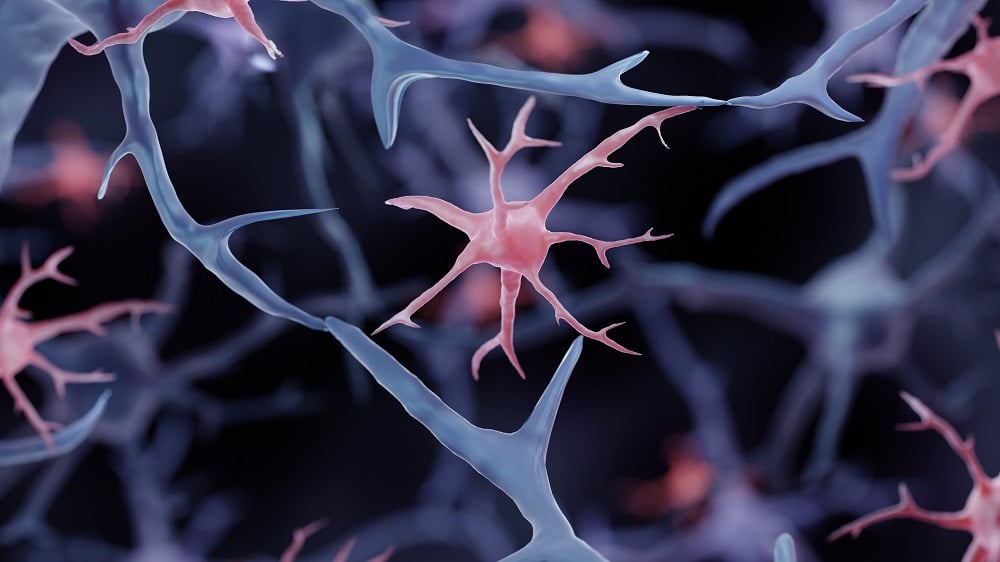
Engineering Microglia to Deliver an Anti-Alzheimer’s Drug
This study represents a proof of principle for employing engineered microglia as vectors for disease-modifying therapies for Alzheimer’s disease.
Published on June 19, 2025, the research illustrated innovative strategies for utilizing the brain’s resident immune cells, microglia, to combat the detrimental effects of amyloid plaques in Alzheimer’s disease.
The Challenge of the Blood-Brain Barrier
The delivery of therapeutic compounds to the brain is complicated by the presence of the blood-brain barrier (BBB), a selective permeability barrier that restricts most compounds from entering brain tissue. This protective mechanism, while crucial in maintaining brain integrity, also complicates the administration of much-needed therapies.
- Failure of the BBB can result in serious neurological complications (Profaci et al., 2020).
- Traditional delivery methods include invasive strategies such as direct injections or viral gene therapy, which can present significant risks (Terstappen et al., 2021).
Given these barriers and risks, the researchers opted for using mature microglia, which have shown promise in previous studies for safely engrafting into brain tissue without the tumorigenic risks associated with neural stem cells (Zong et al., 2015).
Engineering Microglia to Respond to Amyloid Plaques
The researchers developed a novel mouse model that lacked endogenous microglia, leading to the accumulation of amyloid plaques associated with Alzheimer’s disease. They utilized induced pluripotent stem cells (iPSCs) to generate microglia (iMG) that produce neprilysin, an enzyme essential for degrading amyloid beta upon detection of plaques through the CD9 receptor.
The study demonstrated that:
- iMG selectively expressed CD9 in the vicinity of plaques, indicating successful targeting.
- Microglia engineered to secrete neprilysin (sNEP) significantly outperformed those that merely expressed neprilysin on their membranes (NEP).
Findings on Efficacy
The research highlighted several key findings regarding the efficacy of the engineered microglia:
| Measurement | sNEP Microglia | NEP Microglia | Control Human Microglia |
|---|---|---|---|
| Amyloid Clearance Rate | 2x faster | 1.5x faster | Baseline |
| Synaptophysin Levels | Near control levels | No significant effect | Normal |
| GFAP Reduction (Astrogliosis) | Significant reduction | N/A | Elevated |
Reduction of Neuroinflammation
The reduction of amyloid burden in conjunction with improvements in synaptic protein levels indicated the potential for restoring neurological health. Moreover, inflammatory markers showed considerable reductions:
- The levels of interleukins and other inflammatory cytokines returned to near control levels, demonstrating significant anti-inflammatory effects.
- This localized approach showed that widespread engraftment of sNEP-microglia was not essential; targeted injections into the hippocampus and cortex sufficed.
“This novel use of microglia to deliver therapeutic agents demonstrates a promising avenue for Alzheimer’s treatment and highlights the potential for targeted interventions.” – Lead Researcher
Implications for Future Research
This study represents a significant advancement in understanding how engineered microglia could function as a delivery system for neurotherapeutics. However, it is essential to caution that:
- This is an early-stage exploration, primarily conducted in controlled settings with mouse models.
- Further investigations are required to evaluate the safety and effectiveness of iPSC-derived microglia in human applications.
As researchers continue to explore the therapeutic potential of microglia, this work may pave the way for novel treatment modalities in Alzheimer’s disease and other neurodegenerative conditions.
Literature Cited
[1] Profaci, C. P., Munji, R. N., Pulido, R. S., & Daneman, R. (2020). The blood–brain barrier in health and disease: Important unanswered questions. Journal of Experimental Medicine, 217(4), e20190062.
[2] Terstappen, G. C., Meyer, A. H., Bell, R. D., & Zhang, W. (2021). Strategies for delivering therapeutics across the blood–brain barrier. Nature Reviews Drug Discovery, 20(5), 362-383.
[3] Amariglio, N., et al. (2009). Donor-derived brain tumor following neural stem cell transplantation in an ataxia telangiectasia patient. PLoS medicine, 6(2), e1000029.
[4] Prasad, S., et al. (2022). Immune responses and immunosuppressive strategies for adeno-associated virus-based gene therapy for central nervous system disorders: current knowledge and approaches. Human Gene Therapy, 33(23-24), 1228-1245.
[5] Zong, H., Parada, L. F., & Baker, S. J. (2015). Cell of origin for malignant gliomas and its implication in therapeutic development. Cold Spring Harbor Perspectives in Biology, 7(5), a020610.
[6] Chadarevian, J. P., et al. (2024). Therapeutic potential of human microglia transplantation in a chimeric model of CSF1R-related leukoencephalopathy. Neuron, 112(16), 2686-2707.
[7] Oakley, H., et al. (2006). Intraneuronal β-amyloid aggregates, neurodegeneration, and neuron loss in transgenic mice with five familial Alzheimer’s disease mutations: potential factors in amyloid plaque formation. Journal of Neuroscience, 26(40), 10129-10140.
[8] Oakley, H., et al. (2006). Intraneuronal β-amyloid aggregates, neurodegeneration, and neuron loss in transgenic mice with five familial Alzheimer’s disease mutations: potential factors in amyloid plaque formation. Journal of Neuroscience, 26(40), 10129-10140.





Discussion