

Novel Tissue Preservation Method Enables Brain–Body Studies in Animal Research
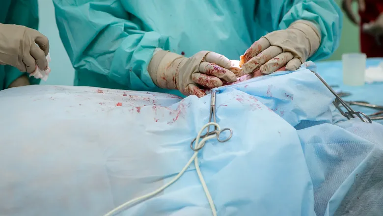
On August 21, 2025, groundbreaking research from cardiovascular scientists at UC Davis Health was published in STAR Protocols. This research unveils a novel method that enables comprehensive studies on the communication between the brain and other vital organs, such as the heart and gut. By advancing traditional techniques, this new approach not only preserves brain tissue but also allows for the simultaneous collection of living (unfixed) samples from various organs during animal research.
The Significance of the New Method
The traditional methodologies employed in neuroscience have typically involved preserving the entire animal body, which precluded the ability to study living tissues from the same organism in detail. With the newly developed dual-preservation technique, researchers can conduct various analyses—including histology and molecular examinations—while using the same animal model.
Xiaodong Zhang, a professor of cardiovascular medicine and the corresponding author of the study, stated,
“This dual-preservation method represents a significant advancement in preclinical research, maximizing the scientific value of each model while reducing the number of animals needed for comprehensive studies.”
Advantages of the Dual-Preservation Method
The empowerment of researchers through this technique presents several notable advantages:
- Maximal Utility: The same animal can now be used for multiple types of studies, leading to richer data collection.
- Cost-Effectiveness: Fewer animals mean lower costs associated with research while ensuring more comprehensive data outcomes.
- Preservation of Fresh Samples: Keeping other organs unfixed allows for dynamic studies on organ interactions in live settings.
Applications in Brain-Body Interaction Studies
The ability to preserve brain tissue while keeping other organs fresh paves the way for extensive research into the complex interactions between the brain and other body systems. This is particularly crucial for understanding conditions that involve cognitive and somatic systems, such as:
| Condition | Impact on Brain-Body Interactions |
|---|---|
| Cardiovascular Diseases | Altered neural regulation contributing to heart health. |
| Diabetes | Changes in brain signaling affecting insulin sensitivity. |
| Gut Disorders | Neural pathways influencing gastrointestinal function. |
Research Findings and Future Directions
This method holds promise not only for understanding the interactions involved in disease processes but also for enhancing therapeutic strategies. The following insights emerge from the current data:
“The dual-preservation method allows us to delve deeper into understanding how the brain and body communicate, which is essential for developing novel treatments for multifaceted diseases.” – Xiaodong Zhang
Future investigations are likely to explore:
- Expanding Models: Investigating additional animal models to diversify research contexts.
- Clinical Applications: Developing protocols for potential adoption in human research contexts.
- Technological Advancements: Integrating cutting-edge imaging techniques for real-time studies on brain-body interactions.
Conclusion
This innovative tissue preservation technique, as described in the work of Phung N. Thai and colleagues, marks a significant leap forward in the realm of preclinical research. By fostering deeper exploration of brain–body communications, it promises to enrich our scientific understanding and treatment possibilities across various health domains.
For more in-depth insights, you may refer to the original article: Novel tissue preservation method enables brain–body studies in animal research (Science X).
References
Phung N. Thai et al, “Protocol for mouse carotid artery perfusion for in situ brain tissue fixation and parallel unfixed tissue collection,” STAR Protocols (2025).
DOI: 10.1016/j.xpro.2025.103699




Discussion