


A revolutionary advancement in microscopy has emerged from Nijmegen, Netherlands, where researchers have unveiled the world's first microscope capable of live imaging biological processes at the nanoscale. Led by Nico Sommerdijk from Radboud University Medical Center, this innovative technique enables the visualization of moving protein complexes, providing unprecedented insight into biological phenomena, including the initial stages of arterial calcification.
Bridging the Gap in Microscopy
Traditionally, researchers faced a significant challenge: they had to choose between examining materials in detail at the molecular level using frozen, motionless samples, or observing living, dynamic materials with reduced detail. The team's breakthrough combines these two approaches, marking a significant leap forward in microscopy technology. This method opens a realm of possibilities for future studies, including the visualization of how the COVID-19 vaccine enters a cell.
Technical Challenges and Solutions
As highlighted by Professor Sommerdijk, "If you want to see protein complexes in such fine detail, you need an electron microscope. But the electron beam used can damage the sample and the surrounding fluid, which is undesirable when you want to observe natural processes in the material over extended periods." To mitigate this issue, researchers developed a technique that includes applying a protective layer around the biological material.
“This opens up various new possibilities for the future, such as visualizing how the COVID-19 vaccine enters a cell or capturing the initial stages of arterial calcification.” – Nico Sommerdijk
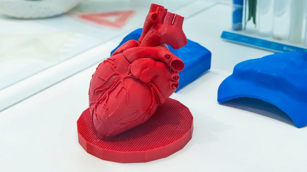
Arterial Calcification Case Study
As a demonstration of this remarkable technique, Sommerdijk and his team focused on arterial calcification, a significant health concern that has yet to be thoroughly understood. The process they established involves wrapping a layer of graphene—a robust material made of a single layer of carbon atoms—around the tissue sample. This action is followed by an immediate freeze to halt biological processes.
After identifying the area of interest with a light microscope, the sample is placed into the electron microscope, where it is warmed up to reactivate biological processes for direct visualization. This innovative workflow enables the observation of complex biochemical interactions on a nanoscale.
Understanding Calcium Phosphate Interactions
Ph.D. candidate Luco Rutten elaborates on the findings: "If there's too much calcium phosphate in the blood, a particular protein in the body can bind to it, preventing it from precipitating. The kidneys then clear it out. Under the microscope, we see that these proteins form small spheres with calcium phosphate, which can still be broken down. However, these spheres can also grow larger, leading to calcified deposits, which can no longer be broken down."
| Finding | Significance |
|---|---|
| Microstructural Visualization | The first evidence of live imaging of protein complexes. |
| Calcium Phosphate Interaction | Insight into the mechanism of arterial calcification. |
| Graphene Application | Effective mitigation of electron beam damage. |
Aiming for the Next Steps
Currently, treatment options for calcified aortic valves are limited to complete valve replacement, as there are no available medications targeting this condition. Sommerdijk stated, "We still don't fully understand what exactly happens with this type of calcification, which is why there are no medications yet." To further investigate this phenomenon, he plans to develop a "heart valve on a chip," a model simulating a healthy heart valve that will introduce calcification mechanisms.
This ambitious project is set to commence in 2025, with the potential to unlock new treatment avenues for cardiovascular diseases associated with calcification.
Conclusion
The advancements made by Sommerdijk and his team signify a crucial step in biological research, particularly in understanding complex processes like arterial calcification. The ability to visualize these processes at the nanoscale in real-time paves the way for significant breakthroughs in both diagnostics and therapeutics.
References
Luco Rutten et al, A Cryo‐to‐Liquid Phase Correlative Light Electron Microscopy Workflow for the Visualization of Biological Processes in Graphene Liquid Cells, Advanced Functional Materials (2024).
Retrieved from Phys.org
Lifespan.io


Discussion