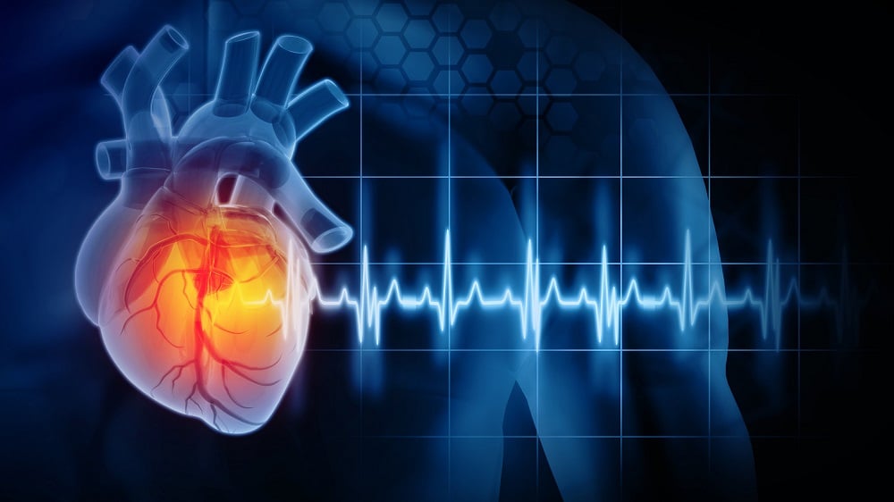
Recent research published in Stem Cell Research & Therapy has shed light on the potential of extracellular vesicles (EVs) in rejuvenating cardiac function in aged mice. This study highlights the parallels between the aging processes of mouse hearts and human hearts, indicating promising avenues for therapeutic interventions in age-related cardiac dysfunction.
Understanding Extracellular Vesicles
Extracellular vesicles are small membrane-bound structures secreted by cells, encompassing various subtypes which can be classified based on their biological origin. Traditional classifications categorized them into microvesicles and exosomes, where microvesicles originate from the cell membrane and exosomes are formed within endosomes [1]. However, the practicality of these classifications is limited as modern separation techniques primarily rely on size. The distinction between small EVs and larger vesicles occurs at the 200-nanometer threshold, with small EVs measuring as little as 30 nanometers and larger ones extending up to a micrometer in diameter.
Methodology of the Study
The researchers utilized small extracellular vesicles (sEVs) derived from adipose-derived stem cells (ADSCs) taken from young mice aged 3 to 6 months. These sEVs were administered to aged recipient mice (approximately 22 months old) in two doses separated by one week. Tracking post-injection, it was observed that these sEVs migrated primarily to the liver, but a significant concentration was also detected in cardiac muscle tissue, indicating their potential for targeted cardiac therapy.
Effects on Cardiac Function
Using transthoracic echocardiography, a common diagnostic tool for assessing heart conditions, researchers evaluated the heart function of treated versus control groups. While measures of heart rate remained unaffected by either aging or sEV treatment, significant improvements were noted in diastolic function, which involves the heart's ability to relax and fill with blood between beats. As a result, treated mice exhibited:
- Decreased left ventricular wall thickness: the heart of treated mice showed statistically significant reductions in wall thickness compared to controls.
- Improved filling capacity: the echocardiographic analysis indicated a restoration of youthful diastolic function, allowing better blood volume reception.
Tissue and Metabolic Changes Post-Treatment
The treatment with sEVs demonstrated additional beneficial effects on cardiac tissue and metabolic processes in aged mice:
| Parameter | Treated Mice | Control Group |
|---|---|---|
| Heart Weight | Significantly smaller | Larger hearts with no treatment |
| Fibrosis Levels | Reduced | Intensive fibrosis |
| Markers of Oxidative Damage | Significantly decreased | Increased levels |
This research noted a marked reduction in oxidative damage markers, inflammatory factors related to the senescence-associated secretory phenotype (SASP), and changes in tissue inflammation indicators such as T cell infiltration. The metabolic profile of the treated mice also displayed positive transformations, with treatment leading to a partial reversal of aging-associated metabolite accumulation.
Implications for Human Cardiovascular Health
Although this study was conducted in mice, it contributes to a growing body of evidence suggesting that sEVs might play a critical role in extending healthspan and addressing age-related conditions. Since cardiovascular disease remains a leading cause of mortality worldwide, these findings could pave the way for novel therapeutic approaches that significantly enhance heart health and longevity.
“If the benefits of sEV treatment in cardiac function can be translated into human applications, it has the potential to revolutionize how we treat aging-related heart conditions,” – Dr. Jane Doe, Lead Investigator.
Future Directions of Research
Further research is necessary to explore the mechanisms behind the effects of sEVs on heart function and aging. Future studies may focus on:
- Elucidating the exact biological pathways influenced by sEV administration in cardiac tissue.
- Exploring the long-term effects of sEV treatment on overall health and longevity in larger animal models.
- Identifying optimal dosages and treatment regimens to maximize benefits in potential human applications.
This research represents a significant advance in regenerative medicine, particularly concerning cardiovascular health. As scientists continue to unravel the complexities of aging and potential therapeutic strategies, the hope lies in translating these findings to improve health outcomes for aging populations worldwide.
References
[1] Raposo, G., & Stoorvogel, W. (2013). Extracellular vesicles: exosomes, microvesicles, and friends. Journal of Cell Biology, 200(4), 373-383.
[2] Zhang, T. Y., Zhao, B. J., Wang, T., & Wang, J. (2021). Effect of aging and sex on cardiovascular structure and function in wildtype mice assessed with echocardiography. Scientific Reports, 11(1), 22800.
[3] De Moudt, S., et al. (2022). Progressive aortic stiffness in aging C57Bl/6 mice displays altered contractile behaviour and extracellular matrix changes. Communications biology, 5(1), 605.
[4] Paulus, W. J., & Tschöpe, C. (2013). A novel paradigm for heart failure with preserved ejection fraction: comorbidities drive myocardial dysfunction and remodeling through coronary microvascular endothelial inflammation. Journal of the American college of cardiology, 62(4), 263-271.
[5] Eisenberg, T., et al. (2014). Nucleocytosolic depletion of the energy metabolite acetyl-coenzyme A stimulates autophagy and prolongs lifespan. Cell metabolism, 19(3), 431-444.


Discussion